
Chinese Bulletin of Botany ›› 2020, Vol. 55 ›› Issue (6): 733-739.DOI: 10.11983/CBB20051 cstr: 32102.14.CBB20051
Previous Articles Next Articles
Fanyu Lin, Xijie Yin*( ), Yuna Liang, Jiechao Huang
Received:2020-03-25
Accepted:2020-07-21
Online:2020-11-01
Published:2020-11-11
Contact:
Xijie Yin
Fanyu Lin, Xijie Yin, Yuna Liang, Jiechao Huang. Analysis of In Situ Distribution of Inorganic Elements in Plants by Micro-XRF[J]. Chinese Bulletin of Botany, 2020, 55(6): 733-739.
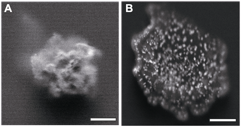
Figure 1 Comparison of fresh sample (A) and gradient dehydrated sample (B) one day after placed at instrument environment Bars=1 mm
| Vacuum (%) | Vented (%) | |||||||
|---|---|---|---|---|---|---|---|---|
| Dwell time (ms) | 200 | 50 | 30 | 10 | 200 | 50 | 30 | 10 |
| Mg | 4.6 | 4.6 | 4.6 | 4.7 | 4.7 | 5.2 | 5.1 | 4.7 |
| Al | 2.3 | 2.4 | 2.3 | 2.3 | 3.6 | 3.1 | 3.6 | 3.9 |
| Si | 1.4 | 1.3 | 1.4 | 1.5 | 0.6 | 0.5 | 0.6 | 0.6 |
| P | 0.7 | 0.7 | 0.8 | 0.8 | 0.2 | 0.2 | 0.2 | 0.3 |
| S | 2.1 | 2.1 | 2.2 | 2.2 | 0.6 | 0.6 | 0.6 | 0.6 |
| K | 13.7 | 13.6 | 13.7 | 13.7 | 12.8 | 12.6 | 12.8 | 12.4 |
| Ca | 38.2 | 38.9 | 39.3 | 39.4 | 36.5 | 36.5 | 37.2 | 36.2 |
| Cr | 3.6 | 3.6 | 3.6 | 3.5 | 3.6 | 3.4 | 3.6 | 3.8 |
| Mn | 3.4 | 3.4 | 3.4 | 3.3 | 3.4 | 3.5 | 3.3 | 3.6 |
| Fe | 11.8 | 11.6 | 11.2 | 11.5 | 14.4 | 14.6 | 13.6 | 13.4 |
| Ni | 1.6 | 1.6 | 1.5 | 1.5 | 2.2 | 2.1 | 2.2 | 2.5 |
| Cu | 2.0 | 2.0 | 1.9 | 1.9 | 2.8 | 2.6 | 2.8 | 2.9 |
| Zn | 1.6 | 1.6 | 1.5 | 1.5 | 2.1 | 2.1 | 2.0 | 2.4 |
| As | 0.5 | 0.4 | 0.4 | 0.4 | 0.8 | 0.5 | 0.6 | 0.7 |
| Cd | 9.1 | 9.0 | 9.0 | 8.7 | 6.8 | 7.2 | 6.6 | 7.0 |
| Pb | 3.3 | 3.3 | 3.1 | 3.1 | 5.1 | 5.3 | 5.2 | 5.0 |
Table 1 Micro-XRF area scanning results under different conditions
| Vacuum (%) | Vented (%) | |||||||
|---|---|---|---|---|---|---|---|---|
| Dwell time (ms) | 200 | 50 | 30 | 10 | 200 | 50 | 30 | 10 |
| Mg | 4.6 | 4.6 | 4.6 | 4.7 | 4.7 | 5.2 | 5.1 | 4.7 |
| Al | 2.3 | 2.4 | 2.3 | 2.3 | 3.6 | 3.1 | 3.6 | 3.9 |
| Si | 1.4 | 1.3 | 1.4 | 1.5 | 0.6 | 0.5 | 0.6 | 0.6 |
| P | 0.7 | 0.7 | 0.8 | 0.8 | 0.2 | 0.2 | 0.2 | 0.3 |
| S | 2.1 | 2.1 | 2.2 | 2.2 | 0.6 | 0.6 | 0.6 | 0.6 |
| K | 13.7 | 13.6 | 13.7 | 13.7 | 12.8 | 12.6 | 12.8 | 12.4 |
| Ca | 38.2 | 38.9 | 39.3 | 39.4 | 36.5 | 36.5 | 37.2 | 36.2 |
| Cr | 3.6 | 3.6 | 3.6 | 3.5 | 3.6 | 3.4 | 3.6 | 3.8 |
| Mn | 3.4 | 3.4 | 3.4 | 3.3 | 3.4 | 3.5 | 3.3 | 3.6 |
| Fe | 11.8 | 11.6 | 11.2 | 11.5 | 14.4 | 14.6 | 13.6 | 13.4 |
| Ni | 1.6 | 1.6 | 1.5 | 1.5 | 2.2 | 2.1 | 2.2 | 2.5 |
| Cu | 2.0 | 2.0 | 1.9 | 1.9 | 2.8 | 2.6 | 2.8 | 2.9 |
| Zn | 1.6 | 1.6 | 1.5 | 1.5 | 2.1 | 2.1 | 2.0 | 2.4 |
| As | 0.5 | 0.4 | 0.4 | 0.4 | 0.8 | 0.5 | 0.6 | 0.7 |
| Cd | 9.1 | 9.0 | 9.0 | 8.7 | 6.8 | 7.2 | 6.6 | 7.0 |
| Pb | 3.3 | 3.3 | 3.1 | 3.1 | 5.1 | 5.3 | 5.2 | 5.0 |
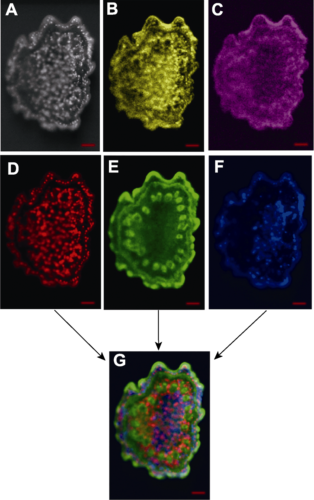
Figure 2 Distribution characteristics of different elements in stem cross section (A) Stem cross section; (B) P; (C) S; (D) Ca; (E) K; (F) Mn; (G) Ca, K, Mn overlay. Bars=500 μm
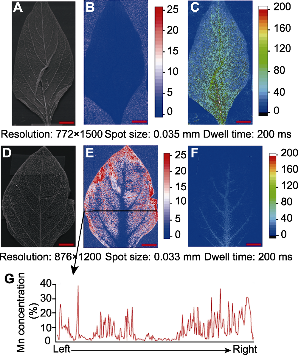
Figure 3 Distribution characteristics of Mn in leaf (A)-(C) Control group ((A) Leaf; (B) Mn concentration heatmap; (C) Ca/Mn colormap); (D)-(G) Experimental group ((D) Leaf; (E) Mn concentration heatmap; (F) Ca/Mn colormap; (G) Linear distribution of Mn). Bars=5 mm
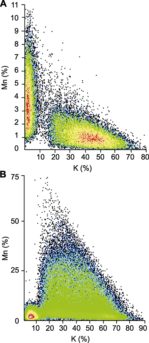
Figure 4 Content characteristics of Mn and K in leaves (A) Control group; (B) Experimental group
| [1] | 陈同斌, 黄泽春, 黄宇营, 谢华, 廖晓勇 (2003). 砷超富集植物中元素的微区分布及其与砷富集的关系. 科学通报 48, 1163-1168. |
| [2] | 丁广大, 刘佳, 石磊, 徐芳森 (2010). 植物离子组学: 植物营养研究的新方向. 植物营养与肥料学报 16, 479-484. |
| [3] | 梁述廷, 刘玉纯, 刘瑱, 林庆文 (2013). X射线荧光光谱微区分析在铅锌矿石鉴定上的应用. 岩矿测试 32, 897-902. |
| [4] | 凌雪, 吴萌蕾, 廖原, 周羿辰 (2018). 文物研究与保护中的无损分析技术. 光谱学与光谱分析 38, 2026-2031. |
| [5] | 罗立强, 沈亚婷, 马艳红, 许涛, 储彬彬, 曾远, 柳检 (2017). 微区X射线荧光光谱仪研制及元素生物地球化学动态分布过程研究. 光谱学与光谱分析 37, 1003-1008. |
| [6] | 沈亚婷 (2014). 原位微区同步辐射X射线荧光和近边吸收谱研究拟南芥幼苗及根际土壤中铅分布与形态特征. 光谱学与光谱分析 34, 818-822. |
| [7] | 王毅, 徐欣, 刘立新 (1994). 生物样品X射线微区分析样品制备技术分析. 植物学通报 11( 专辑), 85, 88. |
| [8] | 许涛, 罗立强 (2011). 原位微区X射线荧光光谱分析装置与技术研究进展. 岩矿测试 30, 375-383. |
| [9] | 余锦涛, 郭占成, 冯婷, 支歆 (2014). X射线光电子能谱在材料表面研究中的应用. 表面技术 43(11), 119-124. |
| [10] | 余志峰, 高永宏, 毛振才 (2005). 植物样品中多种微量元素的分析方法及应用研究. 见: 甘肃省化学会第二十四届年会论文集. 兰州: 甘肃省化学会. pp. 306-308. |
| [11] | Harvey MA, Erskine PD, Harris HH, Brown GK, Pilon-Smits EAH, Casey LW, Echevarria G, van der Ent A (2020). Distribution and chemical form of selenium in Neptunia amplexicaulis from Central Queensland, Australia. Metallomics 12, 514-527. |
| [12] | Modlitbová P, Pořízka P, Kaiser J (2020). Laser-induced breakdown spectroscopy as a promising tool in the elemental bioimaging of plant tissues. TrAC Trends Anal Chem 122, 115729. |
| [13] |
van der Ent A, Przybyłowicz WJ, de Jonge MD, Harris HH, Ryan CG, Tylko G, Paterson DJ, Barnabas AD, Kopittke PM, Mesjasz-Przybyłowicz J (2018). X-ray elemental mapping techniques for elucidating the ecophysiology of hyperaccumulator plants. New Phytol 218, 432-452.
DOI URL PMID |
| [14] |
Wu B, Becker JS (2012). Imaging techniques for elements and element species in plant science. Metallomics 4, 403-416.
URL PMID |
| Viewed | ||||||
|
Full text |
|
|||||
|
Abstract |
|
|||||
 Home
Home