
Chinese Bulletin of Botany ›› 2015, Vol. 50 ›› Issue (1): 72-82.DOI: 10.3724/SP.J.1259.2015.00072 cstr: 32102.14.SP.J.1259.2015.00072
• EXPERIMENTAL COMMUNICATIONS • Previous Articles Next Articles
Qiao Cheng1, 2, Jie Le2, *, Jianguo Zeng1, *
Received:2014-02-17
Accepted:2014-04-03
Online:2015-01-01
Published:2015-04-09
Contact:
Le Jie,Zeng Jianguo
About author:? These authors contributed equally to this paper
Qiao Cheng, Jie Le, Jianguo Zeng. Morphology and Developmental Anatomy of Medicinal Plant Macleaya cordata[J]. Chinese Bulletin of Botany, 2015, 50(1): 72-82.
Add to citation manager EndNote|Ris|BibTeX
URL: https://www.chinbullbotany.com/EN/10.3724/SP.J.1259.2015.00072
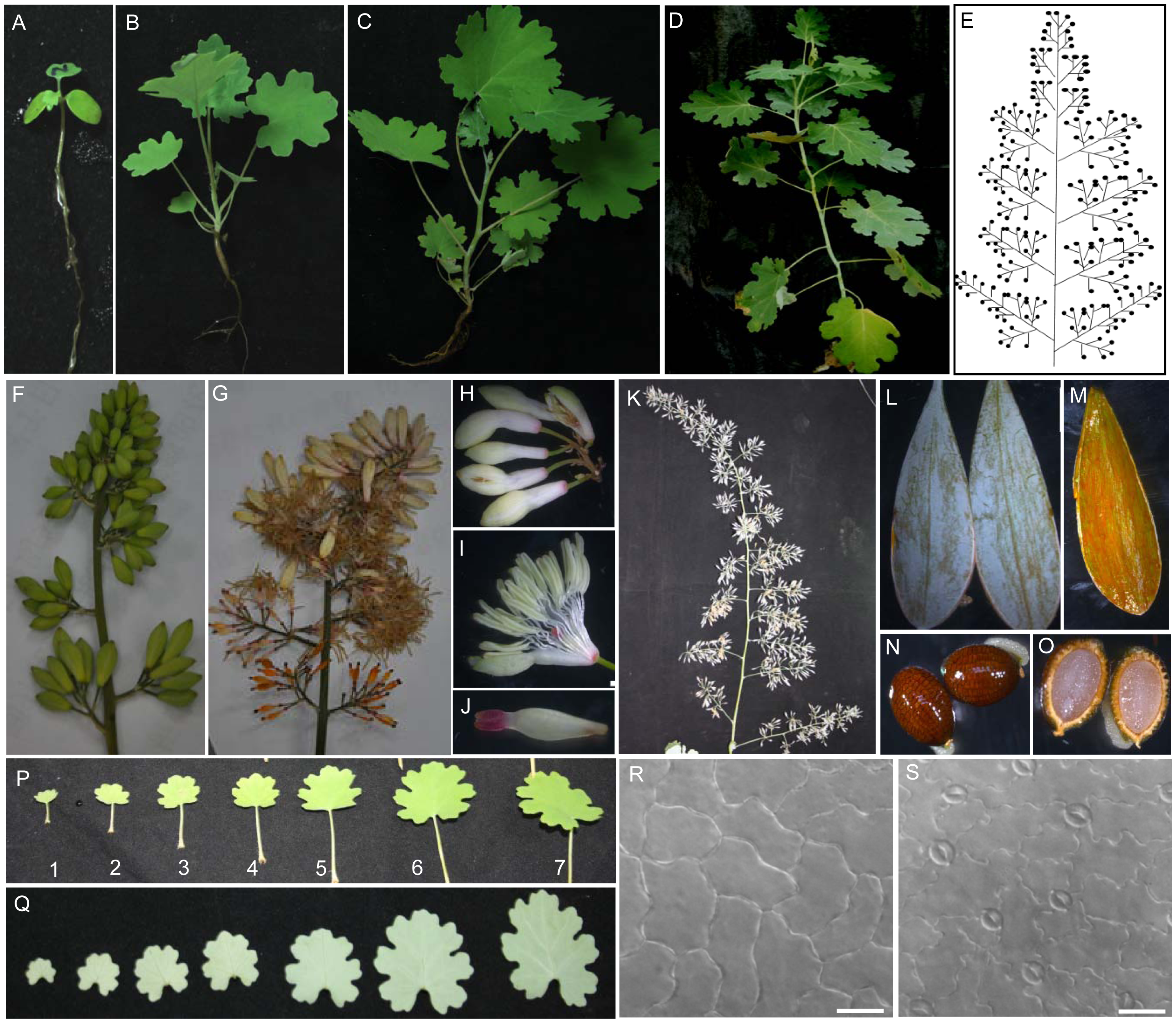
Figure 1 External morphology and leaf epidermis observation of Macleaya cordata (A)-(D) The overall view of one (A), eight (B), twelve (C)- and sixteen (D)-week old plants; (E) Illustration of inflorescence; (F) Part of young inflorescence; (G) Part of mature inflorescence; (H) Flower; (I) Stamens; (J) Pistil; (K) Mature capsule; (L) External morphology of capsule; (M) Internal morphology of capsule; (N) External morphology of seeds; (O) Internal morphology of seeds; (P) Upper surface of leaf one to seven from eight-week-old seedlings; (Q) Lower surface of leaf from one to seven eight-week-old seedlings; (R) Upper epidermis; (S) Lower epidermis. (R), (S) Bar=100 μm

Figure 2 Developmental anatomy of Macleaya cordata roots (A) Seedlings of Macleaya cordata, an arrow points to the position of section; (B) The cross section shows the primary structure of a 4-week-old root, shows the epidermis, cortex and stele; (C) The cross section of a 6-week-old root shows the primary structure to secondary structure; (D) The secondary structure of a 8-week-old root shows the mature metaxylem vessels; (E) The secondary structure of a 10-week-old root shows vascular ray; (F) The secondary structure of a 12-week-old root shows vascular cambium; (G) The secondary structure of a 13-week-old root, showing xylon fiber; (H) The secondary structure of 14-week-old root shows secondary xylem and secondary phloem; (I) The secondary structure of a 16-week-old root shows pericycle; (J) The secondary structure of a 20-week-old root, arrows indicate the crevices in periderm (arrows); Oranges dots in sections are putative accumulation of alkaloids (red arrows). e: Epidermis; c: Cortex; st: Stele; pxy: Primary xylem; pph: Primary phloem; mv: Metaxylem vessels; vr: Vascular ray; vca: Vascular cambium; xf: Xylon fiber; sxy: Secondary xylem; sph: Secondary phloem; pe: Pericycle. (B)-(H) Bar=100 μm; (I) Bar=250 μm; (J) Bar=300 μm
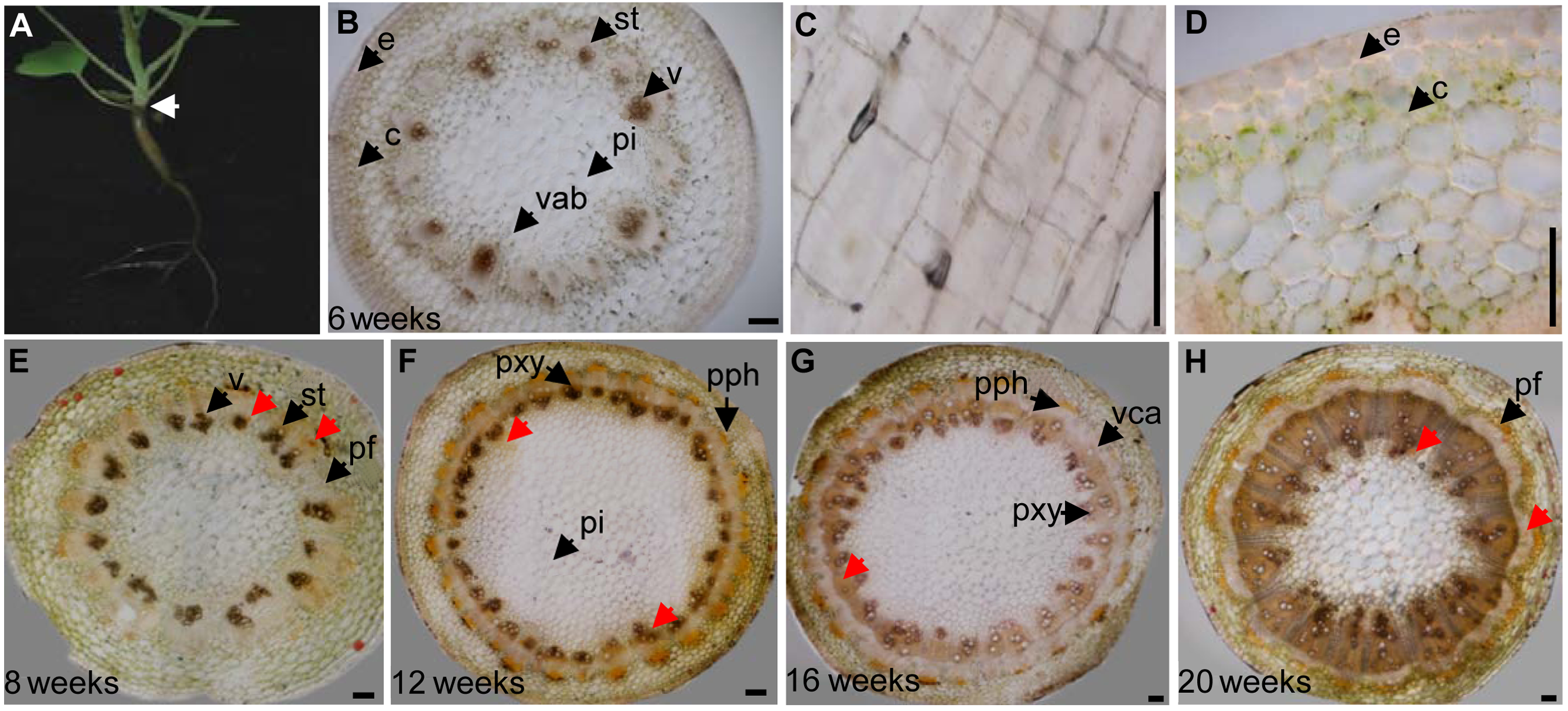
Figure 3 Developmental anatomy of Macleaya cordata stem (A) Part of Macleaya cordata seedlings, an arrow points to the position of section; (B) The cross section of a 6-week-old primary meristem in the stem shows the cortex, epidermis, pith and vascular bundle; (C) Epidermis of stem; (D) Enlarged image shows the epidermis and cortex; (E) The cross section of a 8-week-old primary meristem in the stem shows the differentiation of protoxylem vesseIs and protophloem sieve tubes; (F) The cross section of a 12-week-old stem shows primary xylem, primary phloem and vascular cambium of stem; (G) Secondary structure of a 16-week-old stem shows phloem fiber; (H) The structure of a 20-week-old stem shows vascular ray. Oranges dots in sections are putative accumulation of alkaloids (red arrows). Pi: Pith; pr: Pith ray; e: Epidermis; c: Cortex; vab: Vascular bundle; v: VesseIs; st: Sieve tubes; vca: Vascular cambium; pxy: Primary xylem; pph: Primary phloem; pf: Phloem fiber. (B)-(D) Bar=100 μm; (E)-(H) Bar=200 μm
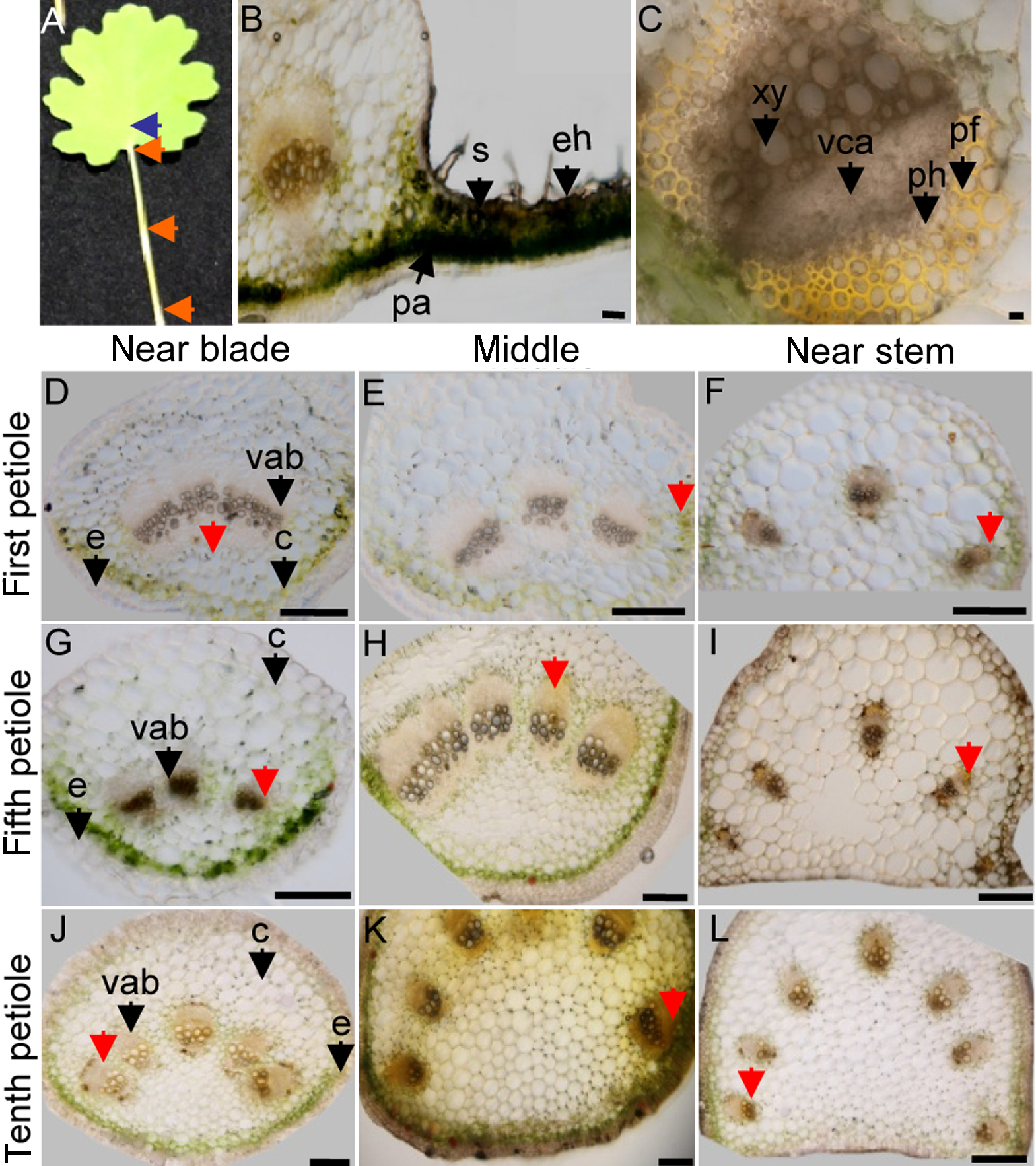
Figure 4 The structure of Macleaya cordata leaf and petiole (A) Image of a leaf; Arrows from bottom to the top point to the positions of section at petioles near blade (panel D, G, J), middle (panel E, H, K), near stem (panel F, I, L) and leaf blade (panel B and C); (B) Palisade and spongy cells in leaves; (C) Vascular bundles include vascular cambium, xylogen, phloem and phloem fiber; (D)-(F) The cross sections of different location on the first petioles; (G)-(I) The cross section of different location on the fifth petioles; (J)-(L) The cross section of different location on the tenth petioles; Orange coloured cells in sections are putative accumulation of alkaloids (red arrows). e: Epidermis; c: Cortex; vab: Vascular bundle; vca: Vascular cambium; pf: Phloem fiber; xy: Xylogen; ph: Phloem; eh: Epidermal hair; pa: Palisade; s: Spongy. (B)-(I) Bar=200 μm; (J) Bar=50 μm; (K) Bar=100 μm; (L) Bar=1 mm
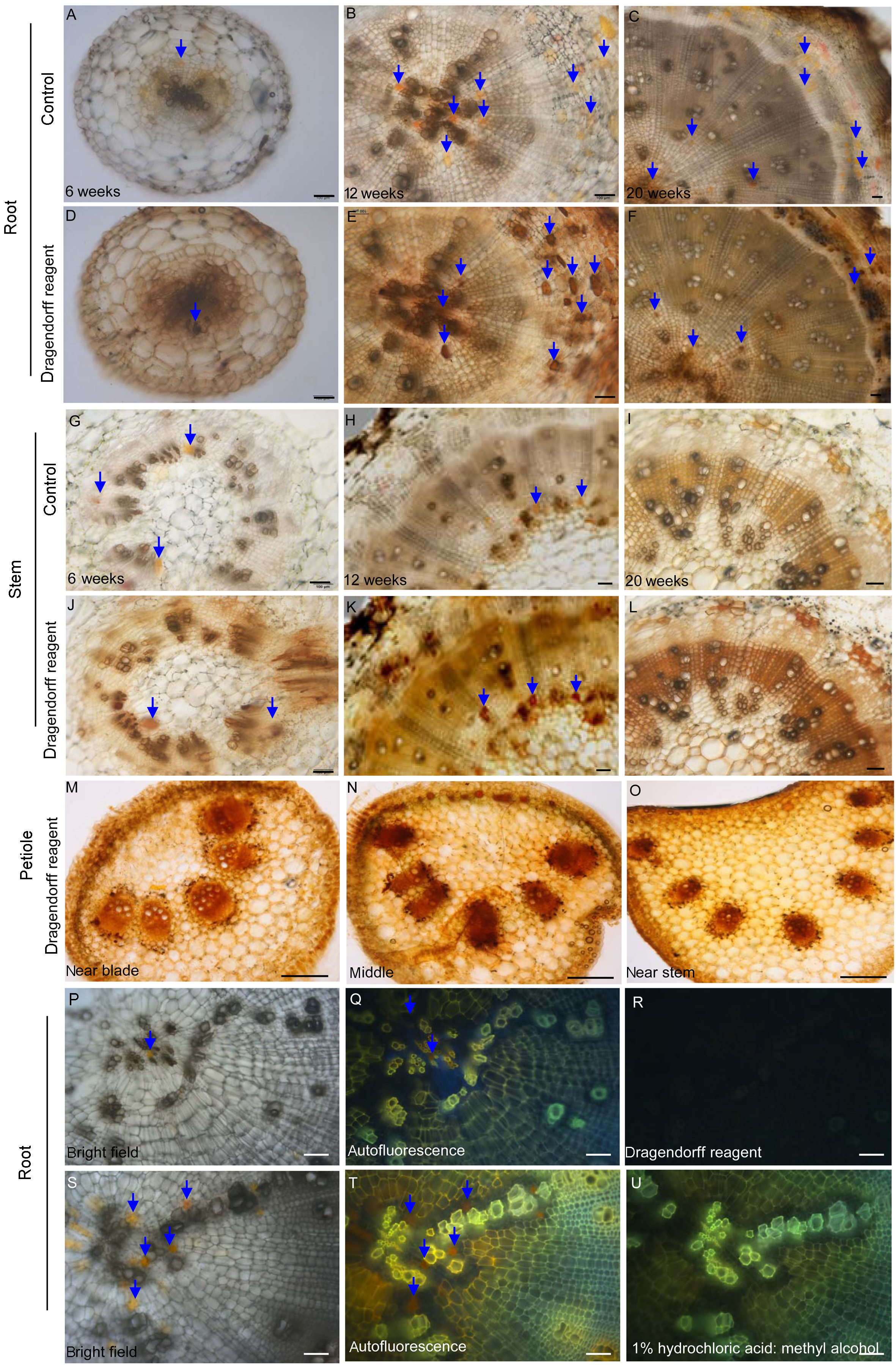
Figure 5 Histochemical localization of alkaloids in Macleaya cordata roots, stems and petioles (A)-(C) Cross sections from 6-, 12-, and 20-week-old roots, respectively; (D)-(F) Images of above root samples stained with Dragendorff reagents; (G)-(I) Cross sections from the 6-, 12-, and 20-week-old stems, respectively; (J)-(L) Images of above stem samples stained with Dragendorff reagents; (M)-(O) The stained sections of a petiole at the position near blade, middle, and near stem; (P)-(R) Alkaloide histochemical localization confirmed by UV-fluorescence; A bright-field image of a root section (P), Fluorescent image after UV stimulation (Q); UV-stimulated fluorescence were not observed after Dragendorff reagent staining (R); (S)-(U) Solvent treatment abolished the UV-stimulated fluorescence; A bright-field image of a root section (S), Image of UV-stimulated fluorescence (T). Fluorescence from alkaloid accumulations were abolished by a treatment with a mixed solvent of 1% hydrochloric acid: methyl alcohol (U). Blue arrows point to the alkaloid accumulations. (A) Bar=100 μm; (B), (C) Bar=200 μm; (D) Bar=100 μm; (E), (F) Bar=200 μm; (G)-(L) Bar=100 μm; (M)-(O) Bar=200 μm; (P)-(U) Bar=100 μm
| 1 | 安彩贤, 杨广德, 叶建涛, 符军梅 (2001). RP-HPLC法同时测定小果博落回中血根碱和白屈菜红碱的含量. 中成药 23, 824-825. |
| 2 | 包英华, 白音, 陈珺霞 (2013). 金钗石斛生物碱的组织化学定位研究. 广西植物 33, 199-202, 190. |
| 3 | 蔡霞, 张爱新, 吴鸿, 胡正海 (1999). 青藤与毛青藤茎中青藤碱积累的组织化学研究. 西北植物学报 19, 104-107. |
| 4 | 陈应庄 (2008). 博落回属植物中苄基异喹啉类生物碱代谢组学研究. 硕士论文. 长沙: 湖南师范大学. |
| 5 | 程辟, 曾建国 (2012). 苯并啡啶类生物碱及其衍生物合成研究进展. 有机化学 32, 1605-1619. |
| 6 | 郭小青, 唐莉苹, 聂建超, 杜海 (2005). 博落回的药理作用及其在动物保健中的应用. 中国动物保健 (5), 34-35. |
| 7 | 郭宇鸽, 曾建国, 谈满良, 黄金红, 刘秀斌 (2011). 博落回叶与小果博落回叶中4种生物碱的含量比较. 中南药学 9, 829-832. |
| 8 | 胡正海 (2010). 植物解剖学. 北京: 高等教育出版社. pp. 215-272. |
| 9 | 黄敬, 曾建国, 罗卫梅, 罗忠勇 (2010). HPLC法同时测定博落回不同部位原阿片碱、别隐品碱的含量. 湖南中医药大学学报 30, 41-42. |
| 10 | 李文亮, 曾建国, 刘发宝, 张雪 (2011). 一测多评法测定博落回果实中原阿片碱、别隐品碱、血根碱和白屈菜红碱的含量. 湖南中医药大学学报 31(7), 25-28. |
| 11 | 李玉萍, 龙绛雪, 曹福祥, 董旭杰 (2010). 蛇根木根部生物碱积累的组织化学定位研究. 中南林业科技大学学报 30(9), 157-161. |
| 12 | 梁彦, 汪矛, 孙小五, 王丽, 郭洁 (2008). 3属4种罂粟科植物幼苗初生维管系统的结构. 植物研究 28, 168-174. |
| 13 | 罗忠勇, 曾建国, 黄敬, 谈满良 (2010). RP-HPLC法测定小果博落回中7种异喹啉类生物碱. 中草药 41, 1188-1190. |
| 14 | 吕洪飞, 罗文姬, 盛仙永, 沈锦波, 毛灵芝 (2006). 二种石斛属植物和小叶石仙桃生物碱的组织化学定位. 中国药学杂志 41, 824-826. |
| 15 | 彭斯文, 张明生, 王玉芳 (2009). 杜鹃兰生物碱组织化学定位初步研究. 世界科学技术-中医药现代化 11, 728-730. |
| 16 | 岐琳, 刘宇凡, 牛晓峰 (2012). 小果博落回的生药学研究. 西北药学杂志 27, 111-112, 127. |
| 17 | 谭玲玲, 廖海民, 蔡霞, 胡正海 (2008). 北柴胡茎、叶的发育解剖学研究. 西北大学学报(自然科学版) 38, 261-266. |
| 18 | 王曙光, 滑培玲, 王涵磊, 胡惠平 (2011). HPLC法测定不同产地博落回中白屈菜红碱和血根碱含量. 安徽医药 15, 434-436. |
| 19 | 魏博菲, 蔡霞, 胡正海 (2011). 泽漆营养器官发育解剖学研究. 西北植物学报 31, 1582-1589. |
| 20 | 杨华中, 黄琼瑶, 刘年猛, 彭飞, 周雪仙, 周天达 (2003). 血水草杀螺成分——血水草生物碱(ECA)的提取. 中国血吸虫病防治杂志 15, 450-451. |
| 21 | 张胜菊, 柯治国, 南玉生 (2003). 博落回抽提物对黄守瓜、菜青虫的田间药效评价. 华中农业大学学报 22, 450-451. |
| 22 | 张遂申, 苏乾元, 刘宏颀 (1989). 中国特有植物血水草属的解剖学研究. 西北植物学报 9, 247-251. |
| 23 | 邹序安, 龙正权 (2009). 博落回的生物学特性与生态价值. 福建林业科技 36, 244-247. |
| 24 | Blank R, Müller-Siegwardt B, Wolffram S (2010). Sang- uinarine does not influence availability or metabolism of tryptophan in pigs.Livest Sci 134, 24-26. |
| 25 | Chen YZ, Liu GZ, Shen Y, Chen B, Zeng JG (2009). Analysis of alkaloids in Macleaya cordata (Willd.) R. Br. using high-performance liquid chromatography with diode array detection and electrospray ionization mass spectrometry.J Chromatogr A 1216, 2104-2110. |
| 26 | Metealfe C, Chalk L (1950). Anatomy of the Dicotyledons. Oxford: Clarendon Press. pp. 74-78. |
| 27 | Samatadze TE, Zelenin AV, Suslina SN, Amosova AV, Popov KV, Zagumennikova TN, Tsytsylin AN, Bykov VA, Muravenko OV (2012). Comparative cytogenetic study of the forms of Macleaya cordata (Willd.) R. Br. from different localities.Genetika 48, 72-79. |
| 28 | Wang GX, Zhou Z, Jiang DX, Han J, Wang JF, Zhao LW, Li J (2010). In vivo anthelmintic activity of five alkaloids from Macleaya microcarpa (Maxim) Fedde against Dactylogyrus intermedius in Carassius auratus.Vet Parasitol 171, 305-313. |
| No related articles found! |
| Viewed | ||||||
|
Full text |
|
|||||
|
Abstract |
|
|||||
 Home
Home