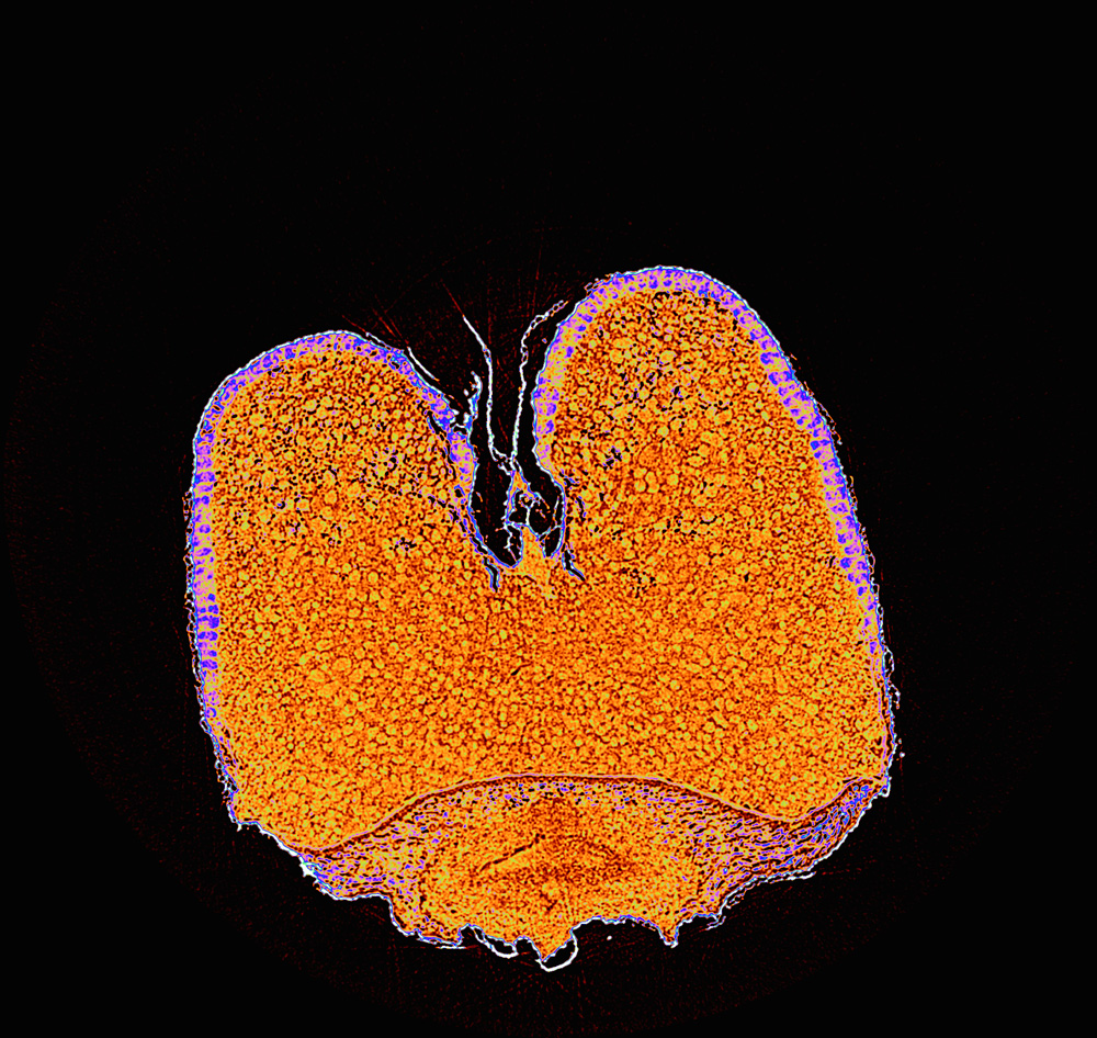

成熟禾谷类作物种子胚乳细胞的显微CT扫描方法
收稿日期: 2024-02-18
录用日期: 2024-03-30
网络出版日期: 2024-04-02
基金资助
中国科学院技术支撑人才项目(2022);黔农科院青年科技基金(2021-20);黔农科院国基后补助(2021-12)
A New Cereal Seed Treatment Method for Displaying Endosperm Cell Structures Under Micro CT Scanning
Received date: 2024-02-18
Accepted date: 2024-03-30
Online published: 2024-04-02
徐秀苹 , 杨小雨 , 冯旻 . 成熟禾谷类作物种子胚乳细胞的显微CT扫描方法[J]. 植物学报, 2025 , 60(1) : 81 -89 . DOI: 10.11983/CBB24022
Cereal starch endosperm is the main source of human staple food, but the methods for observing its mature cell structure are not well developed. Micro CT technology, a non-destructive three-dimensional imaging technique, is a powerful tool for studying plant morphology. However, due to the uniform density of cereal starch endosperm, whose cell structures cannot be distinguished clearly by conventional micro CT techniques. In this study, we used phosphotungstic acid to treat ten different kinds of cereal seeds of seven crops, and then dried them by CO2 critical point dryer. We found that the cell structure of the treated endosperms can be clearly displayed by micro CT. Our method provides a new way for studying the structures and functions of cereal starch endosperm cells.

Key words: micro CT; cereal starch endosperm; phosphotungstic acid
| [1] | Becraft PW, Gutierrez-Marcos J (2012). Endosperm development: dynamic processes and cellular innovations underlying sibling altruism. WIREs Dev Biol 1, 579-593. |
| [2] | Hu J, Guan YJ (2022). Seed Biology, 2nd edn. Beijing: Higher Education Press. pp. 62-63. (in Chinese) |
| 胡晋, 关亚静 (2022). 种子生物学(第2版). 北京: 高等教育出版社. pp. 62-63. | |
| [3] | Huang LJ, Fu WL (2021). A water drop-shaped slingshot in plants: geometry and mechanics in the explosive seed dispersal of Orixa japonica (Rutaceae). Ann Bot 127, 765-774. |
| [4] | Kaestner A, Schneebeli M, Graf F (2006). Visualizing three-dimensional root networks using computed tomography. Geoderma 136, 459-469. |
| [5] | Knipfer T, Reyes C, Earles JM, Berry ZC, Johnson DM, Brodersen CR, McElrone AJ (2019). Spatiotemporal coupling of vessel cavitation and discharge of stored xylem water in a tree sapling. Plant Physiol 179, 1658-1668. |
| [6] | Korte N, Porembski S (2012). A morpho-anatomical characterisation of Myrothamnus moschatus (Myrothamnaceae) under the aspect of desiccation tolerance. Plant Biol 14, 537-541. |
| [7] | Liao H, Fu XH, Zhao HQ, Cheng J, Zhang R, Yao X, Duan XS, Shan HY, Kong HZ (2020). The morphology, molecular development and ecological function of pseudonectaries on Nigella damascena (Ranunculaceae) petals. Nat Commun 11, 1777. |
| [8] | Lin JX, Hu ZJ, Zhang X, Yang SY, Shan XY (2019). Method for Improving Imaging Quality of Arabidopsis thaliana Seeds. Chinese Patent, CN109142398A. 2019-01-04. (in Chinese) |
| 林金星, 胡子建, 张曦, 杨舜垚, 单晓昳 (2019). 一种提高拟南芥种子成像质量的方法. 中国专利, CN109142398A. 2019-01-04. | |
| [9] | Losso A, B?r A, D?mon B, Dullin C, Ganthaler A, Petruzzellis F, Savi T, Tromba G, Nardini A, Mayr S, Beikircher B (2019). Insights from in vivo micro-CT analysis: testing the hydraulic vulnerability segmentation in Acer pseudoplatanus and Fagus sylvatica seedlings. New Phytol 221, 1831-1842. |
| [10] | Perret JS, Al-Belushi ME, Deadman M (2007). Non-destructive visualization and quantification of roots using computed tomography. Soil Biol Biochem 39, 391-399. |
| [11] | Snell P, Wilkinson M, Taylor GJ, Hall S, Sharma S, Sirijovski N, Hansson M, Shewry PR, Hofvander P, Grimberg ? (2022). Characterisation of grains and flour fractions from field grown transgenic oil-accumulating wheat expressing oat WRI1. Plants 11, 889. |
| [12] | Staedler YM, Masson D, Sch?nenberger J (2013). Plant tissues in 3D via X-ray tomography: simple contrasting methods allow high resolution imaging. PLoS One 8, e75295. |
| [13] | Xu XP, Meng SC, Liang RH, Jin WQ, Feng M (2021). New method of improving micro-CT images contrasts on plant samples. J Chin Electron Microsc Soc 40, 460-466. (in Chinese) |
| 徐秀苹, 孟淑春, 梁荣花, 靳婉青, 冯旻 (2021). 磷钨酸和干燥处理提高植物样品显微CT成像对比度的方法. 电子显微学报 40, 460-466. | |
| [14] | Zhang CQ, Li Y (2019). Crop Seed Science, 2nd edn. Beijing: China Agriculture Press. pp. 16-18. (in Chinese) |
| 张春庆, 李岩 (2019). 作物种子学(第2版). 北京: 中国农业出版社. pp. 16-18. | |
| [15] | Zhang X, Hu ZJ, Guo YY, Shan XY, Li XJ, Lin JX (2020). High-efficiency procedure to characterize, segment, and quantify complex multicellularity in raw micrographs in plants. Plant Methods 16, 100. |
/
| 〈 |
|
〉 |