
新疆阿魏特征显微结构的三维原位无损研究
收稿日期: 2017-05-23
录用日期: 2017-10-07
网络出版日期: 2018-09-11
3-D In-situ Non-destructive Structural Characterization of Ferula sinkiangensis
Received date: 2017-05-23
Accepted date: 2017-10-07
Online published: 2018-09-11
刘慧强 , 凯撒·苏来曼 , 孙芸 , 庞渊 , 樊孝喜 , 谢茹 , 柳超 , 段颖妮 , 马燕 . 新疆阿魏特征显微结构的三维原位无损研究[J]. 植物学报, 2018 , 53(3) : 364 -371 . DOI: 10.11983/CBB17104
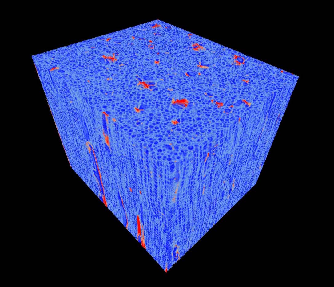
Synchrotron-based X-ray phase-contrast micro-tomography is being used for achieving nondestructive and 3-D characterization due to the high contrast imaging of low Z materials (consisting of C, H, O, N elements). In this paper, we present a new method to combine the high resolution synchrotron-based X-ray phase-contrast imaging technology and phase retrieval algorithm for analyzing and evaluating the 3D inner micro-structures and nondestructive characteristic structures of Ferula sinkiangensis. The method successfully revealed the 3-D micro-structures and characteristics of F. sinkiangensis with high-density resolution, demonstrating that the method is an intuitive and reliable tool of 3D visualization, which has good potential for characterizing and identifying Chinese medicine materials.
| [1] | 黄博, 姜兆玉, 屈红霞, 马三梅 (2010). 龙牙花不同花器官的表皮形态. 植物学报 45, 594-603. |
| [2] | 孔妤, 王忠, 顾蕴洁, 汪月霞 (2008). 植物根内通气组织形成的研究进展. 植物学通报 25, 248-253. |
| [3] | 黎耀东, 付淑媛, 何江, 樊丛照, 李晓瑾 (2016). 新疆特有药用植物新疆阿魏资源现状与分析. 中国现代中药 18, 714-718. |
| [4] | 刘慧强, 任玉琦, 周光照, 和友, 薛艳玲, 肖体乔 (2012). 相移吸收二元性算法用于X射线混合衬度定量显微CT的可行性研究. 物理学报 61, 078701. |
| [5] | 刘慧强, 王玉丹, 任玉琦, 薛艳玲, 和友, 郭瀚, 肖体乔 (2012). 采用吸收修正Bronnikov算法的有机复合样品的X射线显微计算机层析研究. 光学学报 32, 0434001. |
| [6] | 刘家熙, 阎秀峰 (2005). 西藏产四种卷柏科植物的孢子形态观察. 植物学通报 22, 44-49. |
| [7] | 肖体乔, 谢红兰, 邓彪, 杜国浩, 陈荣昌 (2014). 上海光源X射线成像及其应用研究进展. 光学学报 34, 0100001. |
| [8] | 徐晓琴, 倪慧, 魏鸿雁, 贾晓光, 张本刚, 卿德刚 (2013). 新疆地产三种肉苁蓉的显微鉴别研究. 时珍国医国药 24, 881-883. |
| [9] | 薛艳玲, 肖体乔, 吴立宏, 陈灿, 郭荣怡, 杜国浩, 谢红兰, 邓彪, 任玉琦, 徐洪杰 (2010). 利用X射线相衬显微研究野山参的特征结构. 物理学报 59, 5496-5507. |
| [10] | 叶琳琳, 薛艳玲, 倪梁红, 肖体乔 (2014). 种子类中药材的三维显微结构的原位研究. 中国中药杂志 39, 2619-2623. |
| [11] | 叶琳琳, 薛艳玲, 谭海, 陈荣昌, 戚俊成, 肖体乔 (2013). X射线相衬显微层析及其在野山参特征结构的定量三维成像研究. 光学学报 33, 1234002. |
| [12] | Paganin D, Mayo SC, Gureyev TE, Miller PR, Wilkins SW (2002). Simultaneous phase and amplitude extraction from a single defocused image of a homogeneous object.J Microscopy 206, 33-40. |
| [13] | Xie HL, Deng B, Du GH, Fu YN, Chen RC, Zhou GZ, Ren YQ, Wang YD, Xue YL, Peng GY, He Y, Guo H, Xiao TQ (2015). Latest advances of X-ray imaging and biomedical applications beamline at SSRF.Nucl Sci Tech 26, 020102. |
| [14] | Xue Y, Liang Z, Tan H, Ni L, Zhao Z, Xiao T, Xu H (2016). Microscopic identification of Chinese medicinal materials based on X-ray phase contrast imaging: from qualitative to quantitative.J Instrum 11, C07001. |
| [15] | Ye LL, Xue YL, Ni LH, Tan H, Wang YD, Xiao TQ (2013). Application of X-ray phase contrast micro-tomography to the identification of traditional Chinese medicines.J Ins- trum 8, C07006. |
| [16] | Ye LL, Xue YL, Wang YD, Qi JC, Xiao TQ (2016). Identification of ginseng root using quantitative X-ray microtomography.J Ginseng Res 41, 290-297. |
/
| 〈 |
|
〉 |

 首页
首页