
Chloroplast Ultrastructure and Chlorophyll Fluorescence Characteristics of Three Cultivars of Pseudosasa japonica
Received date: 2017-06-10
Accepted date: 2017-10-07
Online published: 2018-09-11
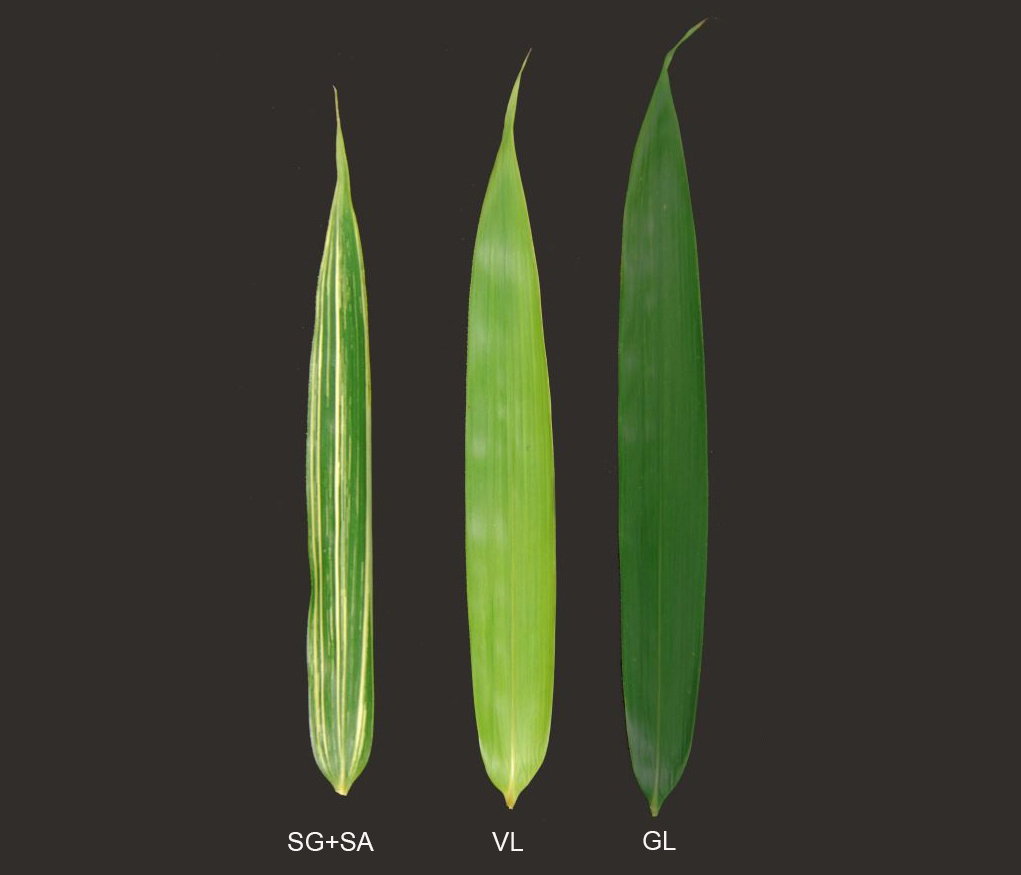
We explored the different photosynthetic characteristics of three Pseudosasa cultivars: P. japonica, P. japonica f. akebonosuji, and P. japonica f. akebono. The differences in photosystem activity and photosynthetic characteristics of different leaf colors were revealed by the changes of chloroplast ultrastructure and fluorescence kinetics curves. The results showed that the photosynthetic pigment content of the three species was significantly different. Except for the intact thylakoid layer structure in the white part of the chloroplast, the green streak and the radix were significantly less than the radix. The chloroplast developmental maturity is inconsistent; the OJIP curve and parameters indicate that the open reduction of the flowering green leaf and the saplings of the PSII reaction center is lower than that of the yam, the capture energy that is used for the electron transfer share to be smaller, and the PSII activity is weaker; The redox balance of the electron transport chain of bamboo leaves P700 to QA is biased towards the reducing side, and it is presumed that the P700 reaction center P700 to PSII primary electron acceptor QA electron transport chain is damaged. Therefore, the chloroplast development caused by changes in PSII activity in the photosystem is immature, which may be the direct cause of the difference in leaf color of the species.

Chen Keyi , Li Zhaona , Cheng Minmin , Zhao Yanghui , Zhou Mingbing , Yang Haiyun . Chloroplast Ultrastructure and Chlorophyll Fluorescence Characteristics of Three Cultivars of Pseudosasa japonica[J]. Chinese Bulletin of Botany, 2018 , 53(4) : 509 -518 . DOI: 10.11983/CBB17115
| 1 | 耿东梅, 单立山, 李毅, Жигунов Анатолий Васильевич (2014). 土壤水分胁迫对红砂幼苗叶绿素荧光和抗氧化酶活性的影响. 植物学报 49, 282-291. |
| 2 | 桂仁意, 刘亚迪, 郭小勤, 季海宝, 贾月, 余明增, 方伟 (2010). 不同剂量137Cs-γ辐射对毛竹幼苗叶片叶绿素荧光参数的影响. 植物学报 45, 66-72. |
| 3 | 何冰, 刘玲珑, 张文伟, 万建民 (2006). 植物叶色突变体. 植物生理学通讯 1, 1-9. |
| 4 | 李鹏民 (2006). 快速叶绿素荧光诱导动力学在植物逆境生理研究中的应用. 博士论文. 泰安: 山东农业大学. pp. 66-73. |
| 5 | 林世青, 许春辉, 张其德, 徐黎, 毛大璋, 匡廷云 (1992). 叶绿素荧光动力学在植物抗性生理学、生态学和农业现代化中的应用. 植物学通报 9, 1-16. |
| 6 | 欧明明, 蔡伟民 (2005). 铁限制对铜绿微囊藻光系统活性变化的影响. 环境化学 24(6), 22-24. |
| 7 | 孙鲁龙, 耿庆伟, 邢浩, 杜远鹏, 翟衡 (2017). 低温处理葡萄根系对叶片PSII活性的影响. 植物学报 52, 159-166. |
| 8 | 唐茜, 施嘉璠 (1997). 川西茶区主栽品种光合强度与叶片结构相关关系的研究. 四川农业大学学报 15(2), 193-198. |
| 9 | 许大全 (2001). 光合作用效率. 上海: 上海科学技术出版社. pp. 136-150. |
| 10 | 杨莉, 郭蔼光, 关旭 (2003). 小麦突变体返白系返白阶段叶绿体超微结构变化研究. 西北农业学报 12(4), 64-67. |
| 11 | 张阿宏, 齐孟文, 张晔晖 (2008). 调制叶绿素荧光动力学参数及其计量关系的意义和公理化讨论. 核农学报 22, 909-912. |
| 12 | 张宪政 (1986). 植物叶绿素含量测定——丙酮乙醇混合液法. 辽宁农业科学 3, 26-28. |
| 13 | 赵云, 王茂林, 李江, 张义正 (2003). 幼叶黄化油菜(Brassica napus L.)突变体Cr3529叶绿体超微结构观察. 四川大学学报(自然科学版) 5, 974-977. |
| 14 | 郑彩霞, 高荣孚 (1999). 光系统I的异质性及其在类囊体膜上分布的研究进展. 北京林业大学学报 21(5), 79-87. |
| 15 | 钟传飞 (2008). 稳态叶绿素荧光动力学理论构建和常绿阔叶植物越冬光合生理生态研究. 博士论文. 北京: 北京林业大学. pp. 47-49. |
| 16 | Deell JR, Murr DP, Wiley L (2003). 1-Methylcyclopropene (1-MCP) increases CO2 injury in apples.Acta Horticul 600, 277-280. |
| 17 | Gilmore AM, Hazlett TL, Govindjee (1997). Xanthophyll cycle-dependent quenching of photosystem II chlorophyll a fluorescence: formation of a quenching complex with a short fluorescence life time.Proc Natl Acad Sci USA 92, 2273-2277. |
| 18 | Heerden PDRV, Strasser RJ, Krüger GHJ (2010). Reduction of dark chilling stress in N2-fixing soybean by nitrate as indicated by chlorophyll a fluorescence kinetics. Physiol Plantarum 121, 239-249. |
| 19 | Lichtenthaler HK (1987). Chlorophylls and carotenoids: pigments of photosynthetic biomembranes.Methods Enzymol 148, 350-382. |
| 20 | Murchie EH, Lawson T (2013). Chlorophyll fluorescence analysis: a guide to good practice and understanding so- me new applications.J Exp Bot 64, 3983-3998. |
| 21 | Papageorgiou GC, Tsimillimichael M, Stamatakis K (2007). The fast and slow kinetics of chlorophyll a fluorescence induction in plants, algae and cyanobacteria: a viewpoint.Photosynth Res 94, 275-290. |
| 22 | Schreiber U (2004). Pulse-amplitude-modulation (PAM) fluo- rometry and saturation pulse method: an overview. In: Papageorgiou GC, Govindjee, eds. Chlorophyll a Fluorescence. Heidelberg: Springer. pp. 279-319. |
| 23 | Strasser RJ, Tsimilli-michael M, Srivastava A (2004). Analysis of the chlorophyll a fluorescence transient. In: Papageorgiou GC, Govindjee, eds. Chlorophyll a Fluorescence: A Signature of Photosynthesis. Dordrecht: Springer. pp. 321-362. |
| 24 | Tsimilli-Michael M, Strasser RJ (2008). In vivo assessment of stress impact on Plant’s Vitality: Applications in Detecting and Evaluating the Beneficial Role of Mycorrhization on Host Plants. In: Varma A, eds. Heidelberg: Springer. pp. 679-703. |
| 25 | Wang Q, Sullivan RW, Kight A, Henry RL, Huang JR, Jones AM, Korth KL (2004). Deletion of the chloroplast- localized Thylakoid formation1 gene product in Arabidopsis leads to deficient thylakoid formation and variegated leaves. Plant Physiol 136, 3594-3604. |
| 26 | Waters MT, Langdale JA (2009). The making of a chloroplast.EMBO J 28, 2861-2873. |
| 27 | Wu JX, Zhang ZG, Zhang Q, Han X, Gu XF, Lu TG (2015). The molecular cloning and clarification of a photorespiratory mutant, oscdm1, using enhancer trapping. Front Ge- net 6, 226. |
| 28 | Zhu XG, Govindjee, Baker NR, deSturler E, Ort DR, Long SP (2005). Chlorophyll a fluorescence induction kinetics in leaves predicted from a model describing each discrete step of excitation energy and electron transfer associated with Photosystem II.Planta 223, 114-133. |
/
| 〈 |
|
〉 |