
冷冻聚焦离子束-扫描电镜成像技术研究进展
收稿日期: 2021-09-14
录用日期: 2021-11-17
网络出版日期: 2021-11-17
基金资助
国家自然科学基金(31801201)
Advances in Cryo-focused Ion Beam-Scanning Electron Microscopy Imaging Technology
Received date: 2021-09-14
Accepted date: 2021-11-17
Online published: 2021-11-17
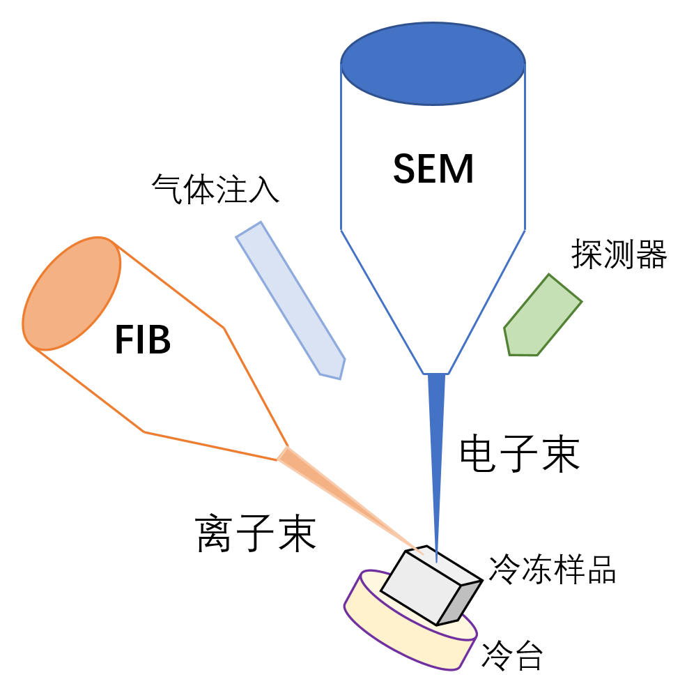
冷冻聚焦离子束-扫描电镜成像(Cryo-FIB-SEM)是一种新兴的成像检测技术, 在原位进行冷冻聚焦离子束切割和冷冻扫描电镜成像, 为研究天然含水状态下生物样品内部未被破坏的原始结构打开了一扇窗口。近年来, 该技术在生命科学领域的应用研究取得了一系列重要进展。该文对其在冷冻体积连续成像、冷冻光电关联成像、冷冻透射扫描成像、冷冻含水切片制备监控及冷冻扫描图像处理等方面的研究进展进行综述, 并展望了该技术在大体积生物样品三维原位成像研究领域的前沿性发展趋势, 以期推动Cryo-FIB-SEM技术在生物样品三维结构研究中的应用。
关键词: 冷冻聚焦离子束-扫描电镜成像; 冷冻光电关联; 冷冻体积连续成像; 冷冻透射扫描成像
贾星 , 孙飞 , 季刚 . 冷冻聚焦离子束-扫描电镜成像技术研究进展[J]. 植物学报, 2022 , 57(1) : 24 -29 . DOI: 10.11983/CBB21161
Cryo-focused ion beam-scanning electron microscopy (Cryo-FIB-SEM) is an emerging technology designed for advanced imaging detection, which performs in situ by combining Cryo-FIB milling and Cryo-SEM imaging, and has facilitated the visualization of the native structures of biological sample in the context of the cellular environment in the frozen hydrated state. In recent years, a series of important advances have been achieved in the application of this technology in the research field of life science. In this review, we summarize its application in cryo-volume serial imaging, and in combination with cryo-correlative light and electron microscopy (CLEM), cryo-transmission SEM (TSEM), cryo-lamella preparation monitoring, and Cryo-SEM image processing. We also provide future prospective on future development and application of Cryo-FIB-SEM in three-dimensional in situ imaging of large volume biological samples.
| [1] | Bhawana, Miller JL, Cahoon AB (2014). 3D Plant cell architecture of Arabidopsis thaliana (Brassicaceae) using focused ion beam-scanning electron microscopy. Appl Plant Sci 2, apps.1300090. |
| [2] | Mesman RJ, Hayles MF, Schneijdenberg CTWM, Mathisen C, Post JA (2013). In-situ integrity control of frozen-hydrated, vitreous lamellas prepared by the cryo-focused ion beam-scanning electron microscope. J Struct Biol 183, 11-18. |
| [3] | Guo Q, Lehmer C, Martínez-Sánchez A, Rudack T, Beck F, Hartmann H, Pérez-Berlanga M, Frottin F, Hipp MS, Hartl FU, Edbauer D, Baumeister W, Fernández-Busnadiego R (2018). In situ structure of neuronal C9orf72 Poly-GA aggregates reveals proteasome recruitment. Cell 172, 696-705. |
| [4] | Hayles MF, De Winter DAM (2021). An introduction to cryo-FIB-SEM cross-sectioning of frozen, hydrated life science samples. J Microsc 281, 138-156. |
| [5] | Hoffman DP, Shtengel G, Xu CS, Campbell KR, Freeman M, Wang L, Milkie DE, Pasolli HA, Iyer N, Bogovic JA, Stabley DR, Shirinifard A, Pang S, Peale D, Schaefer K, Pomp W, Chang CL, Lippincott-Schwartz J, Kirchhausen T, Solecki DJ, Betzig E, Hess HF (2020). Correlative three-dimensional super-resolution and block- face electron microscopy of whole vitreously frozen cells. Science 367, eaaz5357. |
| [6] | Kizilyaprak C, Stierhof YD, Humbel BM (2019). Volume microscopy in biology: FIB-SEM tomography. Tissue Cell 57, 123-128. |
| [7] | Li MJ, Ma JF, Li XM, Sui SF (2021). In situ cryo-ET structure of phycobilisome-photosystem II supercomplex from red alga. eLife 10, e69635. |
| [8] | Lučić V, Rigort A, Baumeister W (2013). Cryo-electron tomography: the challenge of doing structural biology in situ. J Cell Biol 202, 407-419. |
| [9] | Mendonça L, Howe A, Gilchrist JB, Sheng YW, Sun DP, Knight ML, Zanetti-Domingues LC, Bateman B, Krebs AS, Chen L, Radecke J, Li VD, Ni T, Kounatidis I, Koronfel MA, Szynkiewicz M, Harkiolaki M, Martin- Fernandez ML, James W, Zhang PJ (2021). Correlative multi-scale cryo-imaging unveils SARS-CoV-2 assembly and egress. Nat Commun 12, 4629. |
| [10] | Scher N, Rechav K, Paul-Gilloteaux P, Avinoam O (2021). In situ fiducial markers for 3D correlative cryo-fluorescence and FIB-SEM imaging. iScience 24, 102714. |
| [11] | Schertel A, Snaidero N, Han HM, Ruhwedel T, Laue M, Grabenbauer M, Möbius W (2013). Cryo FIB-SEM: volume imaging of cellular ultrastructure in native frozen specimens. J Struct Biol 184, 355-360. |
| [12] | Spehner D, Steyer AM, Bertinetti L, Orlov I, Benoit L, Pernet-Gallay K, Schertel A, Schultz P (2020). Cryo- FIB-SEM as a promising tool for localizing proteins in 3D. J Struct Biol 211, 107528. |
| [13] | Titze B, Genoud C (2016). Volume scanning electron microscopy for imaging biological ultrastructure. Biol Cell 108, 307-323. |
| [14] | Vidavsky N, Akiva A, Kaplan-Ashiri I, Rechav K, Addadi L, Weiner S, Schertel A (2016). Cryo-FIB-SEM serial milling and block face imaging: large volume structural analysis of biological tissues preserved close to their native state. J Struct Biol 196, 487-495. |
| [15] | Wagenknecht T, Hsieh C, Marko M (2015). Skeletal muscle triad junction ultrastructure by focused-ion-beam milling of muscle and cryo-electron tomography. Eur J Transl Myol 25, 4823. |
| [16] | Wagner J, Schaffer M, Fernández-Busnadiego R (2017). Cryo-electron tomography-the cell biology that came in from the cold. FEBS Lett 591, 2520-2533. |
| [17] | Wang Q, Huang Y, Ren Z, Zhang X, Ren J, Su J, Zhang C, Tian J, Yu Y, Gao GF, Li L, Kong Z (2020). Transfer cells mediate nitrate uptake to control root nodule symbiosis. Nat Plants 7, 800-808. |
| [18] | Weiss GL, Kieninger AK, Maldener I, Forchhammer K, Pilhofer M (2019). Structure and function of a bacterial gap junction analog. Cell 178, 374-384. |
| [19] | Wu GH, Mitchell PG, Galaz-Montoya JG, Hecksel CW, Sontag EM, Gangadharan V, Marshman J, Mankus D, Bisher ME, Lytton-Jean AKR, Frydman J, Czymmek K, Chiu W (2020). Multi-scale 3D cryo-correlative microscopy for vitrified cells. Structure 28, 1231-1237. |
| [20] | Xu CS, Hayworth KJ, Lu ZY, Grob P, Hassan AM, García-Cerdán JG, Niyogi KK, Nogales E, Weinberg RJ, Hess HF (2017). Enhanced FIB-SEM systems for large- volume 3D imaging. eLife 6, e25916. |
| [21] | Zhang JG, Zhang DY, Sun L, Ji G, Huang XJ, Niu TX, Xu JS, Ma CY, Zhu Y, Gao N, Xu W, Sun F (2021). VHUT-cryo-FIB, a method to fabricate frozen hydrated lamellae from tissue specimens for in situ cryo-electron tomography. J Struct Biol 213, 107763. |
| [22] | Zhu Y, Sun DP, Schertel A, Ning JY, Fu XF, Gwo PP, Watson AM, Zanetti-Domingues LC, Martin-Fernandez ML, Freyberg Z, Zhang PJ (2021). Serial cryoFIB/SEM reveals cytoarchitectural disruptions in leigh syndrome patient cells. Structure 29, 82-87. |
/
| 〈 |
|
〉 |

 首页
首页