
3D-SIM结构照明超分辨率显微镜实现蛋白质在植物亚细胞器内的定位
? 共同第一作者
收稿日期: 2015-02-16
录用日期: 2015-03-30
网络出版日期: 2015-05-07
基金资助
国家自然科学基金青年基金(No.31200611)和国家自然科学基金(No.31170731)
Using 3D-SIM Structure Illumination Microscope to Localize Proteins in Plant Subcellular Compartments
? These authors contributed equally to this paper
Received date: 2015-02-16
Accepted date: 2015-03-30
Online published: 2015-05-07
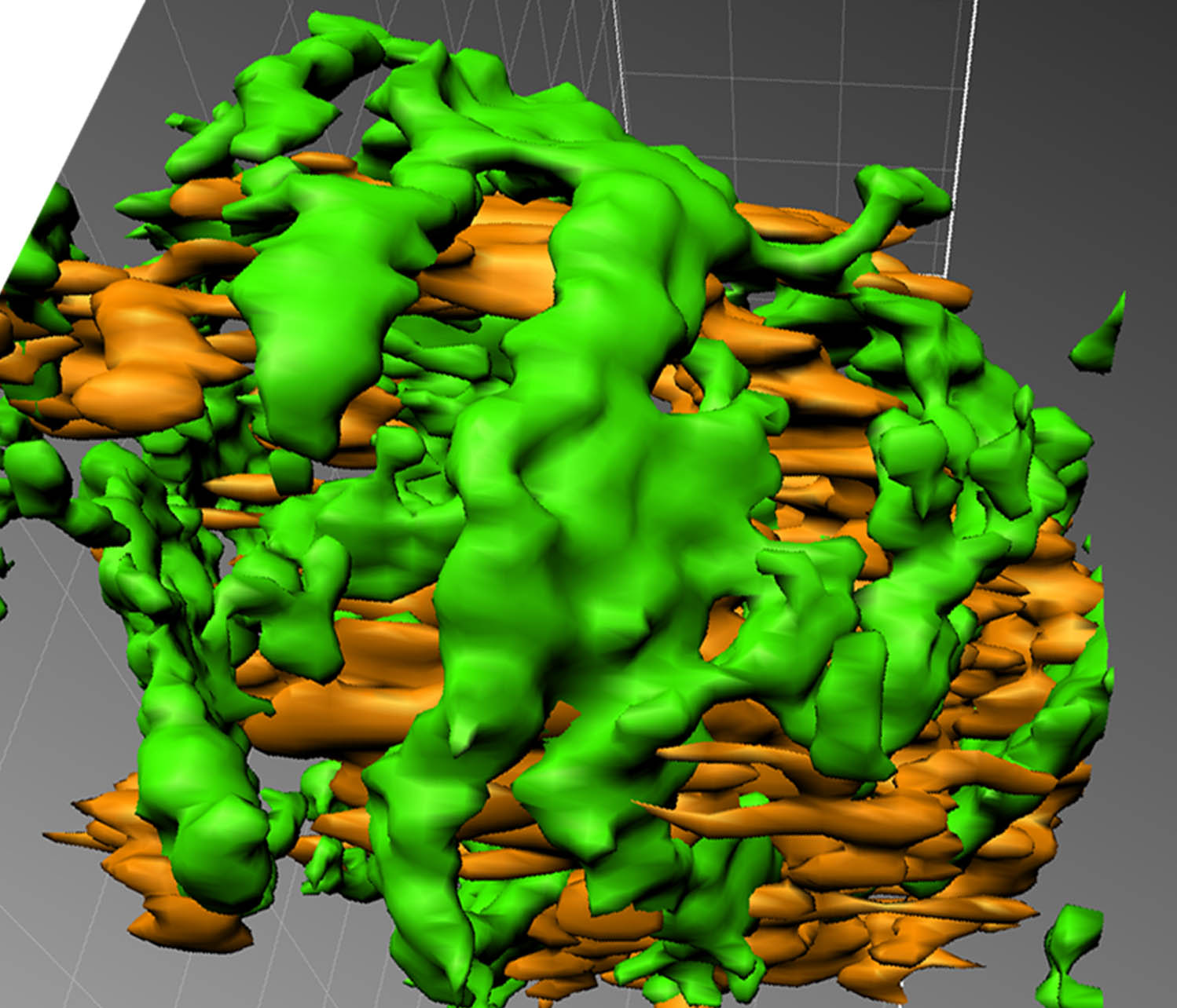
基因表达产物蛋白质的亚细胞定位是解析基因生物学功能的重要证据之一。近年来出现的超分辨率光学成像技术已成功应用于人类和动物细胞中, 预示着显微成像技术继激光共聚焦技术后的又一重要进步。由于植物细胞的特殊性和成像技术的研发取向, 超分辨率光学成像技术在植物细胞蛋白质亚细胞定位的应用尚未见报道。该研究利用DeltaVision OMX显微镜技术, 克服了叶绿体基粒中叶绿素自发荧光与融合蛋白荧光不易区分的缺陷, 解决了受分辨率局限无法将植物细胞中蛋白质在亚细胞器内可视化精确定位的技术难题, 成功地将植物蔗糖合成酶ZmSUS-SH1定位在烟草表皮细胞叶绿体基粒周围。该研究同时建立了一套基于撕片制片法的简便OMX显微镜制片方法, 并针对OMX显微成像技术在植物细胞中蛋白质亚细胞定位的应用进行了讨论。
刘玥, 尹悦佳, 梁重阳, 黄殿帅, 王阳, 刘艳芝, 窦瑶, 冯树丹, 郝东云 . 3D-SIM结构照明超分辨率显微镜实现蛋白质在植物亚细胞器内的定位[J]. 植物学报, 2015 , 50(4) : 495 -503 . DOI: 10.11983/CBB15038
The information on protein subcellular localization is important to elucidate protein function, and imaging technology is one of the important approaches to visualize protein localization. However, conventional microscopy techniques can barely resolve details of subcellular structures mainly because of the auto-fluorescence interference with their limited imaging resolution. In recent years, super-resolution optical imaging technologies have been successfully used in human and animal cell research for their 8-fold higher spacial resolution over laser confocal microscopy. Application of these technologies in plant cells has not been reported, probably due to the peculiarity of plant cells. Here, we report the successful use of DeltaVision OMX microscope technology for visualizing Zea mays sucrose synthase 1 (ZmSUS-SH1) around chloroplast grana in transgenic tobacco epidermal cells. OMX microscopy can overcome the cellular chlorophyll fluorescence interference in imaging fluorescent fusion proteins. We also developed an optimized sample preparation protocol for using DeltaVision OMX microscope technology with plant materials.
| 1 | 龚燕华, 彭小忠 (2010). 蛋白质相互作用及亚细胞定位原理与技术. 北京: 中国协和医科大学出版社. pp. 210-234. |
| 2 | 李楠, 王黎明, 杨军 (1996). 激光共聚焦显微镜的原理和应用. 军医进修学院学报 17, 232-234. |
| 3 | 邢浩然, 刘丽娟, 刘国振 (2006). 植物蛋白质的亚细胞定位研究进展. 华北农学报 21(增刊), 1-6. |
| 4 | 赵启韬, 苗俊英 (2003). 激光共聚焦显微镜在生物医学研究中的应用. 北京生物医学工程 22, 52-54. |
| 5 | Brown ACN, Oddos S, Dobbie IM, Alakoskela JM, Parton RM, Eissmann P, Neil MAA, Dunsby C, French PMW, Davis I, Davis DM (2011). Remodelling of cortical actin where lytic granules dock at natural killer cell immune synapses revealed by super resolution microscopy.PLoS Biol 9, 1-18. |
| 6 | Coltharp C, Xiao J (2012). Superresolution microscopy for microbiology.Cell Microbiol 14, 1808-1818. |
| 7 | Dickerson D (2010). A study of chromatin dynamics during transcription by fluorescence light microscopy. Doctoral Thesis. Dundee: University of Dundee. |
| 8 | Dobbie IM, King E, Parton RM, Carlton PM, Sedat JW, Swedlow JR, Davis I (2011). OMX: a new platform for multimodal, multichannel wide-field imaging. In: Goldmam RD, ed. Live Cell Imaging, 2nd edn. Cold Spring Harbor: Cold Spring Harbor Laboratory Press. pp. 898-909. |
| 9 | Gustafsson MGL (2008). Super-resolution light microscopy goes live.Nat Methods 5, 385-387. |
| 10 | Gustafsson MGL, Shao L, Carlton PM, Wang CJR, Golubovskaya IN, Cande WZ, Agard DA, Sedat JW (2008). Three-dimensional resolution doubling in wide- field fluorescence microscopy by structured illumination.Biophys J 94, 4957-4970. |
| 11 | Masters BR (2010). The development of fluorescence microscopy. eLS 1-9, doi: 10.1002/9780470015902. a0022- 093. |
| 12 | Minden JS, Agard DA, Sedat JW, Alberts BM (1989). Direct cell lineage analysis in Drosophila melanogaster by time-lapse, three-dimensional optical microscopy of living embryos.J Cell Biol 109, 505-516. |
| 13 | Schermelleh L, Carlton PM, Haase S, Shao L, Winoto L, Kner P, Burke B, Cardoso MC, Agard DA, Gustafsson MGL, Leonhardt H, Sedat JW (2008). Subdiffraction multicolor imaging of the nuclear periphery with 3D structured illumination microscopy.Science 320, 1332-1336. |
| 14 | Seabold GK, Wang PY, Petralia RS, Chang K, Zhou A, McDermott MI, Wang YX, Milgram SL, Wenthold RJ (2012). Dileucine and PDZ-binding motifs mediate synaptic adhesion-like molecule 1 (SALM1) trafficking in hippocampal neurons.J Biol Chem 287, 4470-4484. |
| 15 | Swedlow JR (2012). Innovation in biological microscopy: current status and future directions.BioEssays 34, 333-340. |
| 16 | Tanos BE, Yang HJ, Soni R, Wang WJ, Macaluso FP, Asara JM, Tsou MFB (2013). Centriole distal appendages promote membrane docking, leading to cilia initiation.Genes Dev 27, 163-168. |
| 17 | van de Corput MPC, de Boer E, Knoch TA, van Cappellen WA, Quintanilla A, Ferrand L, Grosveld FG (2012). Super-resolution imaging reveals three-dimensional folding dynamics of the β-globin locus upon gene activation.J Cell Sci 125, 4630-4639. |
| 18 | Weil TT, Xanthakis D, Parton R, Dobbie I, Rabouille C, Gavis ER, Davis I (2010). Distinguishing direct from indirect roles for bicoid mRNA localization factors.Deve- lopment 137, 169-176. |
| 19 | Zheng CY, Wang YX, Kachar B, Petralia RS (2011). Differential localization of SAP102 and PSD-95 is revealed in hippocampal spines using super-resolution light microscopy.Commun Integr Biol 4, 104-105. |
/
| 〈 |
|
〉 |

 首页
首页