核糖体图谱技术在植物学研究中的应用
Recent Advances of Ribosome Profiling in Plants
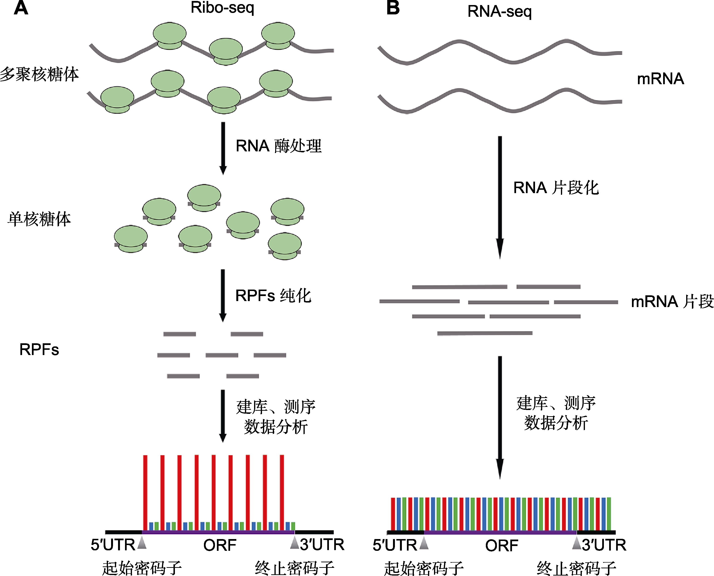
(A) 在核糖体图谱技术(Ribo-seq)中, 多聚核糖体经RNA酶消化成单核糖体, 约30 nt的受核糖体保护的mRNA片段(RPFs)未被RNA酶降解; 将单核糖体解离, 并从中分离和纯化RPFs用于高通量测序, 仅mRNA的蛋白编码区有测序信号; (B) 搭配转录组测序(RNA-seq), 可计算出每条转录本的翻译效率, 在转录组测序中整个转录本上均有测序信号。5′UTR: 5′端非翻译区; 3′UTR: 3′端非翻译区; ORF: 开放阅读框
(A) In the experimental procedure of ribosome profiling (Ribo-seq), polysomes are degraded into monosomes by RNase, resulting in the preservation of the ~30 nucleotides of the ribosome-protected fragments (RPFs) owing to the resistance of the ribosome to the digestion. After isolation and purification from the monosomes, the RPFs are further used for high-throughput sequencing. The ribosome footprints typically show precise positioning within the coding sequence of a mRNA; (B) Ribosome profiling is usually accompanied by a parallel RNA sequencing (RNA-seq) to calculate the translation efficiency of the transcripts. RNA-seq captures mRNA fragments covering the entire transcripts. 5′UTR: 5′ untranslated region; 3′UTR: 3′ untranslated region; ORF: Open reading frame