水稻雄性不育突变体ms102的鉴定和基因定位
王霞1, 严维1, 周志勤2, 常振仪1, 郑敏婷1, 唐晓艳1,2, 吴建新1,*( )
)
)
Identification and Mapping of a Rice Male Sterility Mutant ms102
Xia Wang1, Wei Yan1, Zhiqin Zhou2, Zhenyi Chang1, Minting Zheng1, Xiaoyan Tang1,2, Jianxin Wu1,*()
)
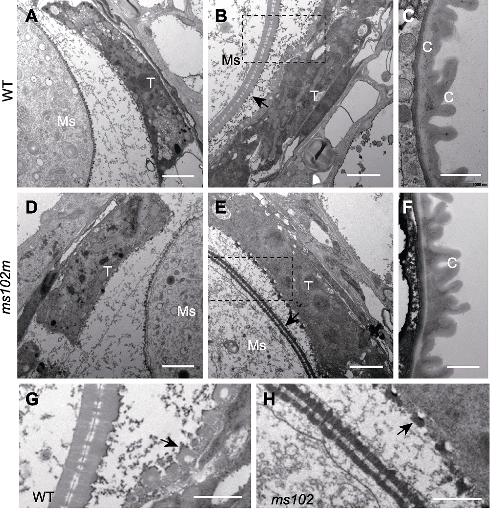
图3. 水稻野生型(WT)和突变体(ms102)花药第9-12时期透射电镜超微结构
(A), (D) 第9时期; (B), (E) 第10时期(箭头指示小孢子外壁); (C), (F) 第12时期; (G), (H) 分别为B和E图中虚线框的放大部分(箭头指示乌氏体); T: 绒毡层; Ms: 小孢子; C: 角质层。(A), (B), (D), (E) Bars=2 μm; (C), (F), (G), (H) Bars =1 μm
Figure 3. Transmission electron microscope images of WT and ms102 anthers at the developmental stages 9-12
(A), (D) Stage 9; (B), (E) Stage 10 (the arrows indicate the microspore wall); (C), (F) Stage 12; (G), (H) The magnified region of dashed box in B and E (the arrows indicate the Ubisch bodies); T: Tapetum; Ms: Microspores; C: Cuticle. (A), (B), (D), (E) Bars=2 μm; (C), (F), (G), (H) Bars=1 μm