)
)
)
)
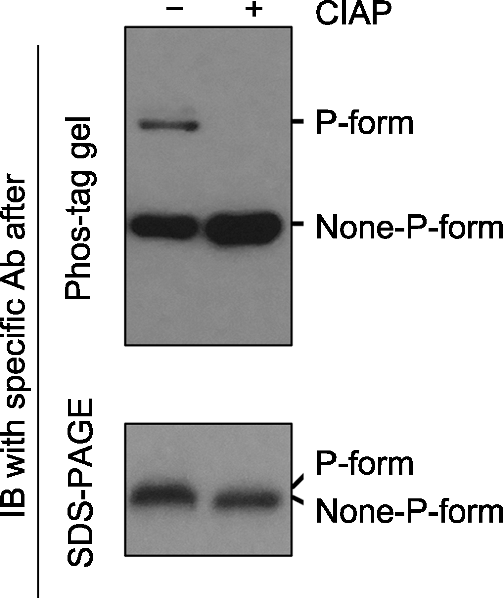
图2. Phos-tag gel和SDS-PAGE gel电泳后, 免疫印迹检测磷酸化蛋白的对比分析
磷酸酶(如Calf intestine alkaline phosphatase, CIAP)可去除磷酸化蛋白上的磷酸基团。同一样品在CIAP处理前(-)、后(+), 磷酸化和非磷酸化形式的蛋白经Phos-tag gel (上图)和SDS- PAGE gel (下图)电泳后的免疫印迹分析结果。经Phos-tag gel电泳, 未加CIAP的样品中特定蛋白的磷酸化(P-form)和非磷酸化形式(None-P-form)被清晰地分开(上图); 加CIAP后磷酸化形式消失而非磷酸化形式条带增强。经SDS-PAGE gel电泳, 未加和加CIAP处理的样品中, 特定蛋白的磷酸化和非磷酸化形式分开不明显(下图)。
Figure 2. Immunoblotting detection of phospho-proteins in samples after Phos-tag gel and SDS-PAGE gel separation
Phospho-proteins can be dephosphorylated by phosphatases (e.g. Calf intestine alkaline phosphatase, CIAP). Protein samples treated with (+) or without (-) CIAP were separated by Phos-tag (top) and SDS-PAGE (bottom) gels, and the specific protein was further detected by immunoblotting. The P-form and None-P-form of the protein were separated clearly by a Phos-tag gel (top), while the two forms were not separated by a SDS-PAGE gel (bottom). The missing of the upper band and the increasing of the bottom band after Phos-tag gel separation indicated that P-form protein was completely dephosphorylated by CIAP treatment.