植物蛋白磷酸化的检测方法
朱丹,曹汉威,李媛,任东涛( )
)
)
Protocols for Analyzing Plant Phospho-proteins
Dan Zhu,Hanwei Cao,Yuan Li,Dongtao Ren()
)
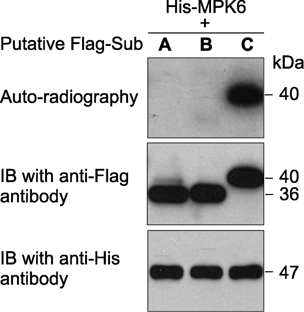
图1. 原核表达的拟南芥激酶MPK6对3个推测底物的体外磷酸分析
激酶(MPK6)连接有6×His标签、推测的底物蛋白(A、B和C)连接有Flag标签。体外磷酸化反应完成后, 反应体系中的蛋白用SDS-PAGE gel分离。然后进行磷酸化底物的放射自显影分析(上图)。底物蛋白(中图)和激酶(下图)的免疫印迹分析显示底物、激酶各自在磷酸化反应中的蛋白使用量基本一致。
Figure 1. An in vitro phosphorylation assay of 3 putative substrate proteins by MPK6 in Arabidopsis thaliana
MPK6 was fused with the 6×His tag and the 3 putative substrates were fused with the Flag tag. After phosphorylation reaction, the proteins in the mixture were separated on a SDS- PAGE gel. The gel was dried and exposed to X-ray film (top). Immunoblotting with anti-His and anti-Flag antibodies were used to show the levels of the substrate (middle) and kinase (bottom) proteins in the reactions.