)
)
)
)
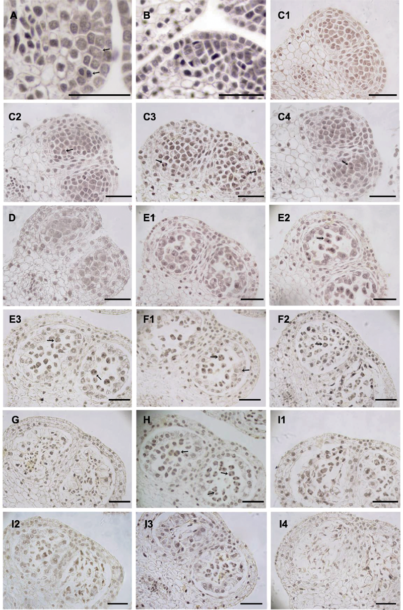
图2. 大叶铁线莲雄花小孢子发育
(A) 孢原细胞(箭头所示, 上)及其平周分裂形成初生壁细胞(箭头所示, 上)和初生造孢细胞(箭头所示, 下); (B) 花药壁; (C1) 次生造孢细胞; (C2) 有丝分裂前期(箭头所示); (C3) 有丝分裂中期(箭头所示, 右)和后期(箭头所示, 左); (C4) 有丝分裂末期(箭头所示); (D) 小孢子母细胞; (E1) 减数第1次分裂前期; (E2) 减数第1次分裂中期(箭头所示); (E3) 减数第1次分裂后期(箭头所示, 右)和末期(箭头所示, 左); (F1) 减数第2次分裂后期(箭头所示, 左), 偶见变形绒毡层(箭头所示, 右); (F2) 减数第2次分裂末期, 偶见左右对称型四分体(箭头所示); (G) 四分体解体; (H) 花药发育异步现象(箭头所示); (I1)-(I4) 小孢子败育。Bars=50 μm
Figure 2. Microspore development in staminat e flower of Clematis heracleifolia
(A) Archesporial cell (arrow, up), primary parietal cell (arrow, up) and primary sporogenous cell (arrow, down); (B) Anther wall; (C1) A row of secondary sporogenous cells; (C2) Prophase (arrow); (C3) Metaphase (arrow, right) and anaphase (arrow, left); (C4) Telophase (arrow); (D) Microspore mother cells; (E1) Microspore mother cells at prophase of prophase I of meiosis; (E2) Microspore mother cells at metaphase of meiosis I (arrow); (E3) Microspore mother cells at anaphase (arrow, right) and telophase (arrow, left) of meiosis I; (F1) Microspore mother cells at anaphase of meiosis II (arrow, left) and amoeboid tapetum (arrow, right); (F2) Microspore mother cells at telophase of meiosis II (arrow) and symmetrical microspore tetrads; (G) Degrading tetrahedral tetrad; (H) The asynchronous phases; (I1)-(I4) Aborted microspores. Bars=50 μm