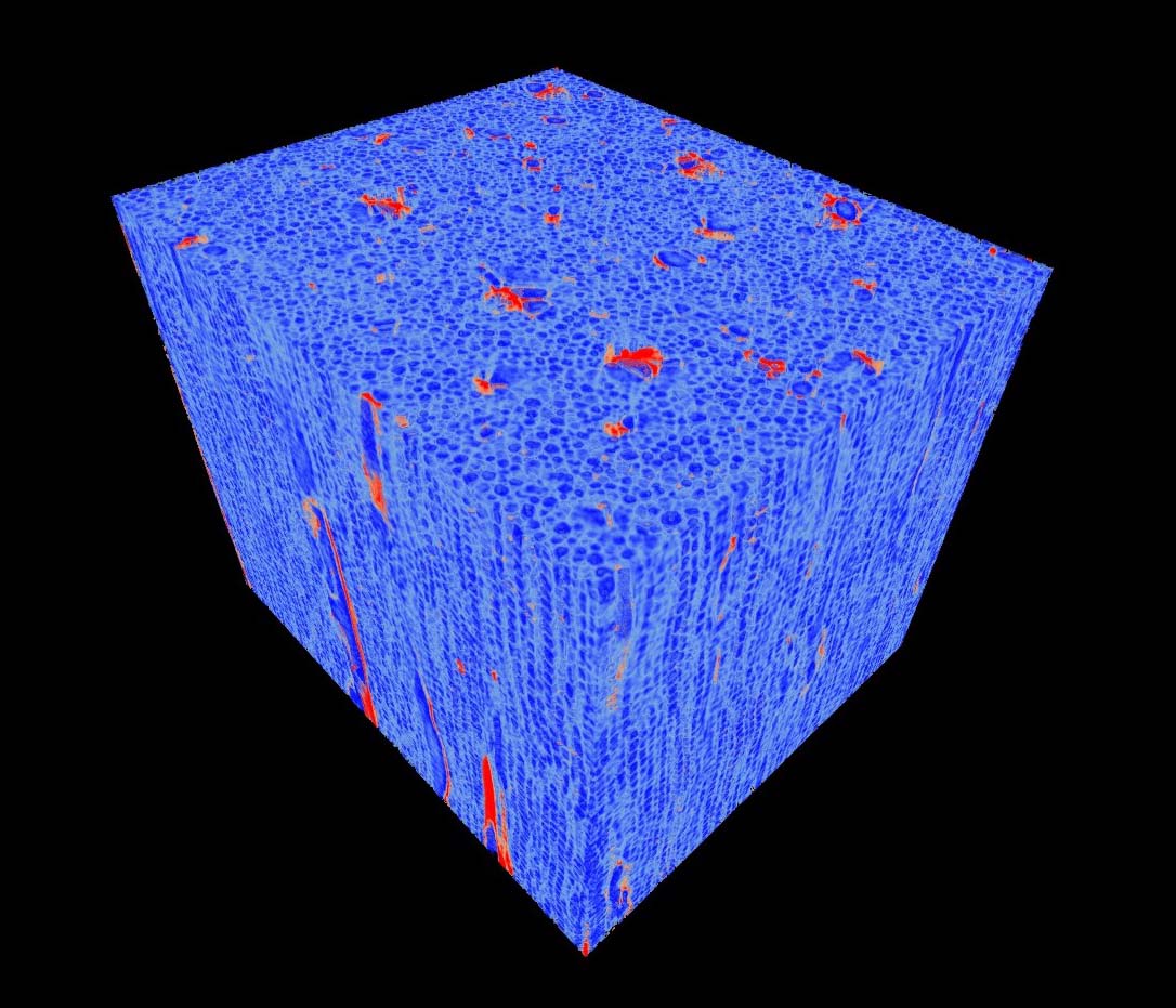
3-D In-situ Non-destructive Structural Characterization of Ferula sinkiangensis
- Liu Huiqiang ,
- Sulaiman·Kaisa ,
- Sun Yun ,
- Pang Yuan ,
- Fan Xiaoxi ,
- Xie Ru ,
- Liu Chao ,
- Duan Yingni ,
- Ma Yan
- 1College of Medical Engineering and Technology, Xinjiang Medical University, Urumqi 830011, China
2Xinjiang Uygur Autonomous Region Institute of Traditional Chinese Medicine and Ethno-Medicine, Urumqi 830002, China
3College of Chinese Medicine, Xinjiang Medical University, Urumqi 830011, China
4Urumqi Hospital of Traditional Chinese Medicine, Urumqi 830000, China
Received date: 2017-05-23
Accepted date: 2017-10-07
Online published: 2018-09-11
Abstract
Synchrotron-based X-ray phase-contrast micro-tomography is being used for achieving nondestructive and 3-D characterization due to the high contrast imaging of low Z materials (consisting of C, H, O, N elements). In this paper, we present a new method to combine the high resolution synchrotron-based X-ray phase-contrast imaging technology and phase retrieval algorithm for analyzing and evaluating the 3D inner micro-structures and nondestructive characteristic structures of Ferula sinkiangensis. The method successfully revealed the 3-D micro-structures and characteristics of F. sinkiangensis with high-density resolution, demonstrating that the method is an intuitive and reliable tool of 3D visualization, which has good potential for characterizing and identifying Chinese medicine materials.

Cite this article
Liu Huiqiang , Sulaiman·Kaisa , Sun Yun , Pang Yuan , Fan Xiaoxi , Xie Ru , Liu Chao , Duan Yingni , Ma Yan . 3-D In-situ Non-destructive Structural Characterization of Ferula sinkiangensis[J]. Chinese Bulletin of Botany, 2018 , 53(3) : 364 -371 . DOI: 10.11983/CBB17104
References
| [1] | 黄博, 姜兆玉, 屈红霞, 马三梅 (2010). 龙牙花不同花器官的表皮形态. 植物学报 45, 594-603. |
| [2] | 孔妤, 王忠, 顾蕴洁, 汪月霞 (2008). 植物根内通气组织形成的研究进展. 植物学通报 25, 248-253. |
| [3] | 黎耀东, 付淑媛, 何江, 樊丛照, 李晓瑾 (2016). 新疆特有药用植物新疆阿魏资源现状与分析. 中国现代中药 18, 714-718. |
| [4] | 刘慧强, 任玉琦, 周光照, 和友, 薛艳玲, 肖体乔 (2012). 相移吸收二元性算法用于X射线混合衬度定量显微CT的可行性研究. 物理学报 61, 078701. |
| [5] | 刘慧强, 王玉丹, 任玉琦, 薛艳玲, 和友, 郭瀚, 肖体乔 (2012). 采用吸收修正Bronnikov算法的有机复合样品的X射线显微计算机层析研究. 光学学报 32, 0434001. |
| [6] | 刘家熙, 阎秀峰 (2005). 西藏产四种卷柏科植物的孢子形态观察. 植物学通报 22, 44-49. |
| [7] | 肖体乔, 谢红兰, 邓彪, 杜国浩, 陈荣昌 (2014). 上海光源X射线成像及其应用研究进展. 光学学报 34, 0100001. |
| [8] | 徐晓琴, 倪慧, 魏鸿雁, 贾晓光, 张本刚, 卿德刚 (2013). 新疆地产三种肉苁蓉的显微鉴别研究. 时珍国医国药 24, 881-883. |
| [9] | 薛艳玲, 肖体乔, 吴立宏, 陈灿, 郭荣怡, 杜国浩, 谢红兰, 邓彪, 任玉琦, 徐洪杰 (2010). 利用X射线相衬显微研究野山参的特征结构. 物理学报 59, 5496-5507. |
| [10] | 叶琳琳, 薛艳玲, 倪梁红, 肖体乔 (2014). 种子类中药材的三维显微结构的原位研究. 中国中药杂志 39, 2619-2623. |
| [11] | 叶琳琳, 薛艳玲, 谭海, 陈荣昌, 戚俊成, 肖体乔 (2013). X射线相衬显微层析及其在野山参特征结构的定量三维成像研究. 光学学报 33, 1234002. |
| [12] | Paganin D, Mayo SC, Gureyev TE, Miller PR, Wilkins SW (2002). Simultaneous phase and amplitude extraction from a single defocused image of a homogeneous object.J Microscopy 206, 33-40. |
| [13] | Xie HL, Deng B, Du GH, Fu YN, Chen RC, Zhou GZ, Ren YQ, Wang YD, Xue YL, Peng GY, He Y, Guo H, Xiao TQ (2015). Latest advances of X-ray imaging and biomedical applications beamline at SSRF.Nucl Sci Tech 26, 020102. |
| [14] | Xue Y, Liang Z, Tan H, Ni L, Zhao Z, Xiao T, Xu H (2016). Microscopic identification of Chinese medicinal materials based on X-ray phase contrast imaging: from qualitative to quantitative.J Instrum 11, C07001. |
| [15] | Ye LL, Xue YL, Ni LH, Tan H, Wang YD, Xiao TQ (2013). Application of X-ray phase contrast micro-tomography to the identification of traditional Chinese medicines.J Ins- trum 8, C07006. |
| [16] | Ye LL, Xue YL, Wang YD, Qi JC, Xiao TQ (2016). Identification of ginseng root using quantitative X-ray microtomography.J Ginseng Res 41, 290-297. |